Ciencia y Salud, Vol. 10, No. 3, septiembre-diciembre, 2026 • ISSN (impreso): 2613-8816 • ISSN (en línea): 2613-8824
MEDIAN RAPHE CYST ASSOCIATED WITH GRADE III PHIMOSIS IN A FIVE-YEAR-OLD BOY: A CASE REPORT
Quiste del rafe medio asociado con fimosis grado III en un niño de cinco años: reporte de caso
DOI: https://doi.org/10.22206/cisa.2026.v10i3.3732
Freddy Alexander Aldaz Vallejo1*, Wendy Vanessa Vaca Vega2, Diana Elizabeth Parrales Suárez3, Cesar Geovany Gia Correa4
Received: 13/04/2026 ● Accepted: 30/04/2026

How to cite: Aldaz Vallejo, F. A., Vaca Vega, W. V., Parrales Suárez, D. E., Gia Correa, C. G. (2026). Median raphe cyst associated with grade III phimosis in a five-year-old boy: a case report. Ciencia y Salud, 10(3), 109-114. https://doi.org/10.22206/cisa.2026.v10i3.3732
Abstract
Background: Median raphe cyst is an uncommon benign congenital lesion related to abnormal embryologic development of the male genital midline. In children, it may be asymptomatic and mimic other cystic lesions.
Case presentation: A five-year-old boy evaluated for grade III phimosis had an elongated, soft, translucent cystic lesion on the perineal/penoscrotal median raphe, without urinary symptoms or signs of infection. Ultrasonography showed a fluid-filled lesion of approximately 10 cc with no urethral communication. Complete excision was performed under general anesthesia, with circumcision during the same procedure. Histopathology confirmed a median raphe cyst lined by pseudostratified/cuboidal epithelium with mucinous goblet cells. At four-month follow-up, healing was satisfactory and no recurrence was observed. Conclusion: Clinical examination, ultrasonography, and histopathology supported definitive single-stage management of this rare pediatric lesion.
Keywords: Median raphe cyst, phimosis, circumcision, ultrasonography, case report.
Resumen
Introducción: El quiste del rafe medio es una lesión congénita benigna infrecuente derivada de alteraciones del desarrollo embrionario de la línea media genital. En niños puede ser asintomático y confundirse con otras lesiones quísticas.
Presentación del caso: Niño de cinco años evaluado por fimosis grado III, en quien se identificó una lesión quística alargada, blanda y translúcida en el rafe medio perineal/penoescrotal, sin síntomas urinarios ni signos de infección. La ecografía evidenció una lesión líquida de aproximadamente 10 cc, sin comunicación uretral. Se realizó exéresis completa y circuncisión en el mismo acto quirúrgico. La histopatología confirmó quiste del rafe medio revestido por epitelio pseudoestratificado/cúbico con células caliciformes mucinosas. A los cuatro meses no hubo recurrencia. Conclusión: La evaluación clínica, ecográfica e histopatológica permitió tratamiento definitivo y seguro en un solo tiempo.
Palabras clave: Quiste del rafe medio, fimosis, circuncisión, ultrasonografía, reporte de caso.
Introduction
Median raphe cyst is a rare benign developmental lesion of the male genitalia that may occur along the ventral midline from the urethral meatus and penile shaft to the scrotum, penoscrotal raphe, perineum, and anus. Its embryologic relevance is linked to abnormal fusion of the urethral folds and sequestration of urethral or epithelial tissue during genital development, which may explain the heterogeneous epithelial lining described on histopathology1-5.
The condition is clinically important because it may be overlooked in childhood, remain asymptomatic, or be confused with other midline genital and perineal cystic lesions. Small, stable, asymptomatic lesions may be observed with clinical follow-up; however, complete excision is generally preferred when the lesion is symptomatic, enlarging, cosmetically concerning, infected, or diagnostically uncertain3, 5-8.
This report describes a perineal/penoscrotal median raphe cyst identified during evaluation of grade III phimosis in a five-year-old boy. The case highlights the value of careful genital and perineal examination, preoperative ultrasonography to exclude urethral communication, and single-stage surgical management when a concomitant foreskin indication requires operative treatment.
Case presentation
Patient information and clinical findings
A five-year-old boy was evaluated in the pediatric surgery outpatient clinic for follow-up of grade III phimosis. During physical examination, an elongated cystic lesion was identified on the perineal/penoscrotal median raphe, in the ventral midline region. The lesion was smooth, translucent, soft, and non-tender, without erythema, discharge, or signs of local infection (Figure 1). No urinary symptoms were reported.
Figure 1. Preoperative appearance of the lesion. Elongated cystic lesion located along the perineal/penoscrotal median raphe
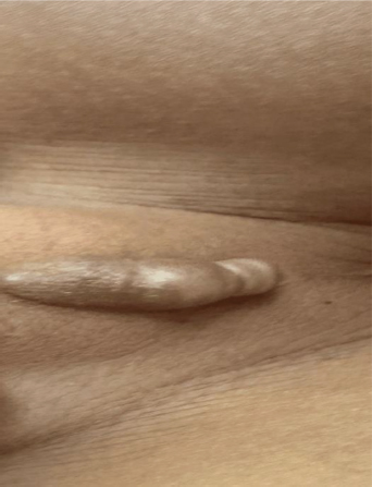
The exact linear length of the cyst was not documented in the available clinical record. The objective size estimate available for this report was the ultrasonographic volume, which was approximately 10 cc.
Diagnostic assessment
Because of the midline location and cystic appearance, ultrasonography was requested for preoperative assessment. Imaging demonstrated a fluid-filled lesion with an estimated volume of approximately 10 cc and no communication with the urethra. These findings supported the diagnosis of a benign cystic lesion and were useful for operative planning.
The differential diagnosis included epidermoid or dermoid cyst, parameatal urethral cyst, preputial cyst, urethral diverticulum, lymphatic malformation, and other benign midline perineal lesions. Absence of urethral communication on ultrasonography reduced concern for urethral involvement.
Therapeutic intervention
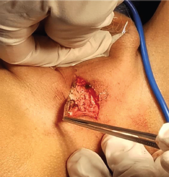
The patient underwent surgery under general anesthesia. Intraoperatively, the cystic lesion was carefully dissected from the surrounding perineal/penoscrotal raphe tissues and completely excised while preserving adjacent structures (Figure 2). Because of the associated grade III phimosis, circumcision was performed during the same procedure as definitive management of the foreskin abnormality. The immediate postoperative course was uneventful, and the patient was discharged in stable condition.
Figure 2. Intraoperative resection of the median raphe cyst. Complete surgical excision of the ventral perineal/penoscrotal cystic lesion
Histopathology, follow-up, and outcomes
Histopathological examination revealed a localized cyst within the dermis, without communication with the epidermis, lined by pseudostratified columnar or cuboidal epithelium with mucinous goblet cells. These findings were consistent with a median raphe cyst.
At four-month follow-up, the patient remained asymptomatic, with satisfactory wound healing and no clinical evidence of recurrence. A formal patient perspective was not obtained because the patient was a young child; no postoperative concerns were recorded from the legal guardian during follow-up. The main clinical, diagnostic, therapeutic, and follow-up events are summarized chronologically in Table 1.
Timeline
Table 1. Case timeline according to CARE reporting structure
Clinical moment |
Main finding or intervention |
Outpatient assessment |
Grade III phimosis and elongated cystic lesion on the perineal/penoscrotal median raphe. |
Preoperative imaging |
Ultrasonography: fluid-filled lesion of approximately 10 cc; no communication with the urethra. |
Surgery |
Complete cyst excision under general anesthesia; circumcision performed in the same operative time. |
Pathology |
Median raphe cyst lined by pseudostratified/cuboidal epithelium with mucinous goblet cells. |
Four-month follow-up |
Asymptomatic patient; adequate healing; no clinical recurrence. |
Discussion
Median raphe cyst is an uncommon congenital lesion of the male genital and perineal midline. Its presentation in childhood may be incidental, particularly when another genital condition prompts examination. In the present case, the lesion was identified during evaluation of grade III phimosis, which influenced operative planning and allowed both conditions to be treated during one anesthetic event.
The perineal/penoscrotal location is relevant because median raphe lesions can occur anywhere along the embryologic fusion line, from the meatus to the perineum. This distribution supports the proposed mechanisms of incomplete urethral fold fusion and epithelial sequestration during development. Histopathological confirmation was essential in this patient because the epithelial lining with pseudostratified/cuboidal cells and mucinous goblet cells falls within the recognized spectrum of median raphe cysts1, 3-5.
Management of asymptomatic median raphe cysts is individualized. Observation with follow-up may be appropriate for small, stable, asymptomatic lesions, especially when there is no diagnostic uncertainty or functional concern. Conversely, complete excision is the most definitive approach for enlarging, symptomatic, infected, cosmetically concerning, or diagnostically uncertain cysts, and it reduces the likelihood of recurrence related to incomplete removal3, 5-8. In this case, excision was favored because the lesion was relatively large by ultrasonographic volume, located in the perineal/penoscrotal raphe, and was being managed during the same operative session as the phimosis.
The coexistence of grade III phimosis also influenced treatment choice. Current evidence supports topical corticosteroids with gentle retraction as first-line treatment for many boys with symptomatic phimosis, while surgical intervention, including circumcision, is considered for persistent or pathological phimosis, recurrent inflammatory complications, failed conservative treatment, or when concomitant operative management is clinically justified10-12. In the present patient, single-stage circumcision and cyst excision provided definitive treatment while avoiding a second anesthetic procedure.
Preoperative ultrasonography was useful because it confirmed the cystic nature of the lesion, provided the available objective size estimate, and excluded urethral communication. These aspects are important in the differential diagnosis of parameatal urethral cysts, urethral diverticula, epidermoid or dermoid cysts, lymphatic malformations, and other benign midline perineal lesions5, 8, 9.
This report was structured according to the CARE case report recommendations, including patient information, clinical findings, diagnostic assessment, therapeutic intervention, follow-up, outcomes, and ethical transparency13. Its main limitation is that it describes a single patient and therefore does not allow generalization of findings. In addition, follow-up was limited to four months; although this period documented early healing and absence of short-term recurrence, longer surveillance would be necessary to assess late outcomes.
Conclusions
Median raphe cyst should be considered in the differential diagnosis of pediatric cystic lesions located along the ventral genital or perineal midline. In this case, careful examination during evaluation of grade III phimosis allowed recognition of a perineal/penoscrotal median raphe cyst. Ultrasonography helped define the lesion and exclude urethral communication, histopathology confirmed the diagnosis, and complete excision combined with circumcision provided favorable short-term results.
Funding
No funding was received for this study.
Authors’ contributions
Conceptualization: Freddy Aldaz, Wendy Vaca, Diana Parrales, Cesar Gia.
Investigation: Freddy Aldaz, Wendy Vaca.
Methodology: Diana Parrales, Cesar Gia.
Project administration: Diana Parrales, Cesar Gia.
Writing – original draft: Freddy Aldaz, Wendy Vaca.
Writing – review and editing: Freddy Aldaz, Wendy Vaca.
Ethical compliance and transparency statements
Ethics committee statement
This manuscript corresponds to a single anonymized clinical case. In accordance with current institutional regulations, it was not considered necessary to submit it for formal ethics committee review. If required by the journal or the institution, the authors will process the request for submission to the competent ethics committee.
Informed consent statement
Written informed consent for publication of the case details and clinical images was obtained from the patient’s legal guardian.
Reporting guideline statement
This case report was prepared in accordance with the CARE guidelines.
Use of artificial intelligence
Generative artificial intelligence assistance was used for language editing, formatting, and editorial support. The authors reviewed and verified all scientific content and accept full responsibility for the final manuscript.
Data availability statement
All relevant clinical information is contained within the manuscript.
Conflicts of interest
The authors declare no conflicts of interest.
References
1. Krauel L, Tarrado X, Garcia-Aparicio L, Lerena J, Suñol M, Rodó J, et al. Median raphe cysts of the perineum in children. Urology. 2008;71(5):830-1. https://doi.org/10.1016/j.urology.2007.11.131.
2. Bayne CE, DeMarco RT, Wehry MA. Median raphe cyst of the perineum. J Pediatr. 2020;216:236. https://doi.org/10.1016/j.jpeds.2019.07.050.
3. Syed MMA, Amatya B, Sitaula S. Median raphe cyst of the penis: a case report and review of the literature. J Med Case Rep. 2019;13(1):214. https://doi.org/10.1186/s13256-019-2133-5.
4. Shao IH, Chen TD, Shao HT, Chen HW. Male median raphe cysts: serial retrospective analysis and histopathological classification. Diagn Pathol. 2012;7:121. https://doi.org/10.1186/1746-1596-7-121.
5. Nowowiejska J, Piccolo V, Van Gysel D, Grimalt R, Cutrone M. Diagnosis and management of penile median raphe cysts: a comprehensive review. Dermatol Pract Concept. 2025;15(3):5288. https://doi.org/10.5826/dpc.1503a5288.
6. Matsuyama S, Matsui F, Yazawa K, Matsumoto F, Shimada K, Matsuoka K. Long-term follow-up of median raphe cysts and parameatal urethral cysts in male children. Urology. 2017;101:99-103. https://doi.org/10.1016/j.urology.2016.10.020.
7. Sinopidis X, Athanasopoulou M, Kourea E. Median raphe cysts: why surgical excision should be performed in childhood. J Pediatr. 2021;235:301-2. https://doi.org/10.1016/j.jpeds.2021.04.072.
8. Tanaka K, Nakazawa-Tanaka N, Urao M. Do parameatal urethral cysts in children need surgical excision? Urology. 2022;167:198-200. https://doi.org/10.1016/j.urology.2022.05.026.
9. Elebute OA, Komolafe MD, Akinjo AO, Alakaloko FM. Preputial cyst in the newborn: a case report from Africa. Afr J Paediatr Surg. 2023;20(1):80-2. https://doi.org/10.4103/ajps.ajps_142_21.
10. Moreno G, Ramirez C, Corbalán J, Peñaloza B, Morel Marambio M, Pantoja T. Topical corticosteroids for treating phimosis in boys. Cochrane Database Syst Rev. 2024;1(1):CD008973. https://doi.org/10.1002/14651858.CD008973.pub3.
11. European Association of Urology. EAU Guidelines on Paediatric Urology [Internet]. Arnhem: EAU Guidelines Office; 2025 [citado 2026 Apr 29]. Disponible en: https://uroweb.org/guidelines/paediatric-urology.
12. Dave S, Afshar K, Braga LH, Anderson P. CUA guideline on the care of the normal foreskin and neonatal circumcision in Canadian infants. Can Urol Assoc J. 2018;12(2):E76-E99. https://doi.org/10.5489/cuaj.5033.
13. Gagnier JJ, Kienle G, Altman DG, Moher D, Sox H, Riley D; CARE Group. The CARE guidelines: consensus-based clinical case reporting guideline development. J Med Case Rep. 2013;7:223. https://doi.org/10.1186/1752-1947-7-223.
_______________________________
* Corresponding author: Freddy Alexander Aldaz Vallejo, freddyaldaz6@gmail.com
1 Pediatric Surgeon, Rosales National Hospital, El Salvador. ORCID: https://orcid.org/0009-0003-1586-7731, email: freddyaldaz6@gmail.com
2 General Surgeon, Rosales National Hospital, El Salvador. ORCID: https://orcid.org/0009-0001-8861-532X, email: wendyva7@hotmail.com
3 Pediatric Surgeon, Rosales National Hospital, El Salvador. ORCID: https://orcid.org/0009-0000-7762-8638, email: dradianaparrales@gmail.com
4 Pediatric Surgeon, Hospital Clínica Rodriguez, Ecuador. ORCID: https://orcid.org/0000-0002-3063-4616, email: cgtauro_85@hotmail.com