Ciencia y Salud, Vol. 10, No. 2, mayo-agosto, 2026 • ISSN (impreso): 2613-8816 • ISSN (en línea): 2613-8824
GUT MICROBIOTA AND CARDIOVASCULAR DISEASE: IMPLICATIONS FOR PUBLIC HEALTH
Microbiota intestinal y enfermedades cardiovasculares: implicaciones para la salud pública
DOI: https://doi.org/10.22206/cisa.2026.v10i2.3521
Received: 01/01/2025 • Accepted: 03/07/2025

How to cite: Sauce Guevara, M. A., Guerra Martínez, A. A. (2026). Gut microbiota and cardiovascular disease: implications for public health. Ciencia y Salud, 10(2), 103-117. https://doi.org/10.22206/cisa.2026.v10i2.3521
Abstract
In recent years, the gut microbiota has gained prominence in various physiopathology’s, particularly in cardiovascular diseases (CVD), the leading cause of global mortality. The interaction between bacterial metabolites, systemic inflammation, and regulatory mechanisms of lipid metabolism and endothelial function has generated increasing interest. This narrative review synthesizes current evidence on the relationship between gut microbiota and the development/progression of CVD, with emphasis on compounds such as trimethylamine N-oxide (TMAO) and short-chain fatty acids (SCFAs), as well as the impact of dietary or probiotic interventions. Additionally, we analyze the implications for public health prevention, considering the potential of the microbiota as a risk biomarker and an intervention tool in population-wide strategies.
Keywords: SCFAs, cardiovascular diseases, gut microbiota, public health, TMAO.
Resumen
En los últimos años, la microbiota intestinal ha cobrado protagonismo en diversas patologías fisiológicas, especialmente en las enfermedades cardiovasculares (ECV), principal causa de mortalidad a nivel mundial. La interacción entre los metabolitos bacterianos, la inflamación sistémica y los mecanismos reguladores del metabolismo lipídico y la función endotelial ha generado un interés creciente. Esta revisión narrativa sintetiza la evidencia actual sobre la relación entre la microbiota intestinal y el desarrollo/progresión de las ECV, con énfasis en compuestos como el N-óxido de trimetilamina (TMAO) y los ácidos grasos de cadena corta (AGCC), así como el impacto de las intervenciones dietéticas o probióticas. Además, analizamos las implicaciones para la prevención en salud pública, considerando el potencial de la microbiota como biomarcador de riesgo y herramienta de intervención en estrategias poblacionales.
Palabras clave: AGCC, enfermedades cardiovasculares, microbiota intestinal, salud pública, TMAO.
Introduction
Cardiovascular diseases (CVD) are the leading cause of global mortality, responsible for approximately 17.9 million deaths annually1. Although traditional risk factors such as hypertension, diabetes, and smoking have been extensively studied, in recent years the gut microbiota has emerged as a crucial component in the pathophysiology of CVD2. This microbial ecosystem, essential for metabolic and immunological homeostasis, influences not only digestive health but also systemic processes such as chronic inflammation and lipid metabolism, which are directly linked to CVD3. Recent research indicates that gut dysbiosis, an imbalance in microbial composition, may promote atherosclerosis, hypertension, and heart failure through multiple mechanisms4. Notable among these are the production of pro-inflammatory metabolites like trimethylamine N-oxide (TMAO), associated with increased risk of cardiovascular events, and the reduction of short-chain fatty acids (SCFAs), which exert protective effects on the endothelium and inflammation5–6. This duality underscores the complexity of the microbiota-heart interaction and its potential as a therapeutic target.
Given the growing evidence, it is imperative to critically review the most recent scientific advances to explore innovative strategies such as dietary modulation, the use of probiotics/prebiotics, or even fecal microbiota transplantation (FMT). The gut microbiota has been proposed as a cardiovascular risk biomarker, offering opportunities for personalized interventions and evidence-based public health policies7. This narrative review synthesizes key findings in this field while highlighting diverse perspectives on molecular mechanisms and translational implications with public health impact, thereby guiding future research and clinical applications.
Materials and methods
A narrative review of the scientific literature was conducted. The bibliographic search covered the period 2004–2024 and was executed between January and March 2025 using the following databases: PubMed/MEDLINE, Scopus, Web of Science, and Google Scholar. Key terms in Spanish and English were combined with Boolean operators: ("Gut microbiota" OR "Microbiota") AND ("Cardiovascular diseases" OR "Cardiovascular diseases") AND ("Public health" OR "Public health") AND ("TMAO" OR "Trimethylamine N-oxide") AND ("SCFAs" OR "Short-chain fatty acids" OR "SCFAs").
Results
Gut microbiota: concept and core functions
The gut microbiota constitutes a dynamic ecosystem comprising trillions of microorganisms. These organisms colonize the intestinal niche, an anaerobic and nutritionally favorable environment8. This interaction facilitates a complex symbiotic relationship between commensal microorganisms and their human host9. This microbial community, known as the gut microbiota, includes not only bacteria but also archaea, viruses, and fungi; the collective genetic material of this consortium is termed the gut microbiome4. In humans, the gut microbiota is dominated by bacteria, with diversity exceeding 1,000 species. It is primarily composed of bacteria from the phyla Firmicutes (60–80%), Bacteroidetes (15–30%), Actinobacteria, Proteobacteria, which play crucial roles in digestion, synthesis of vitamins (such as K and B12), and immune system modulation5,10–12. The Firmicutes and Bacteroidetes phyla dominate the large intestine, constituting approximately 90% of the total microbiota. Their ratio serves as an indicator of an individual's health status13. However, this microbial equilibrium is significantly disrupted in various pathologies, particularly cardiovascular diseases (CVD), where dysregulation of the Firmicutes/Bacteroidetes relationship not only reflects pathological states but may also function as a specific diagnostic biomarker14. The microbiota is not a static system. It constantly interacts with the host organism through symbiotic or competitive relationships, thereby regulating key processes, from digestion and immune responses to chronic disease risk15. Ongoing research continues to reveal its critical role as a mediator between nutrition, environment, and human health12.
Factors modulating the gut microbiota
The composition and functionality of the gut microbiota are governed by complex interactions between exogenous and endogenous factors. These factors are detailed below12,16–17:
Diet
Diet represents one of the most significant environmental determinants shaping the gut microbiota. This is exemplified in studies such as De Filippo et al., which contrasted the microbiota of Italian adolescents (consuming a Western diet high in animal fats and protein) with rural African children (whose diet consisted primarily of fiber and complex carbohydrates with minimal animal fats/proteins)12, 18. Analysis revealed substantial differences, supporting the premise that diet profoundly shapes microbial communities. Similarly, studies in healthy adults confirm that sustained dietary patterns (e. g., predominant consumption of simple carbohydrates, fats, or proteins) influence bacterial enterotypes19. The Western diet correlates with microbiota imbalances and increased disease predisposition, whereas alternatives like the Mediterranean diet associate with extended lifespan and reduced cardiometabolic disorders20. Furthermore, food additives such as emulsifiers and artificial sweeteners may disrupt microbial ecology and promote pathology21–22. Collectively, these findings underscore that diet—irrespective of age—modulates gut microbiota composition and influences non-communicable disease risk.
Antibiotics
Modern lifestyles in developed nations, characterized by improved hygiene and excessive antibiotic use (particularly during infancy), disrupt intestinal bacterial colonization. These alterations correlate with increased allergic diseases and autoimmune disorders23. Clinical studies demonstrate a direct correlation between antibiotic exposure frequency (especially broad-spectrum agents) and elevated asthma risk in adolescence24. Animal models reveal that low-dose penicillin in neonates transiently alters microbiota, inducing pathological weight gain and adult obesity predisposition25. These findings indicate that antibiotics interfere with normal microbial colonization, disrupting immunometabolic homeostasis and increasing susceptibility to chronic diseases in adulthood.
Age
Aging modifies the structural and functional activity of the gut microbiota, influencing health status and longevity16. Comparative analyses show increased Bacteroidetes abundance and decreased Firmicutes in older adults (>65 years), accompanied by heightened interindividual heterogeneity26–27. These variations associate with dysregulation of key bacterial metabolites (including SCFAs, vitamins B7/B12, and creatine) implicated in sarcopenia and frailty syndromes28. Nutritional habits critically modulate age-related microbiota changes: geriatric individuals with high-fiber diets exhibit greater α-diversity and attenuated proinflammatory cytokine profiles (TNF-α, IL-6), contrasting sharply with nursing home residents showing Coprobacillus and Parabacteroides predominance29. Notably, centenarians display a distinctive microbial signature enriched in Bifidobacterium, Christensenellaceae, and Akkermansia, suggesting their potential role in longevity30. These data establish gut microbiota as a critical regulator of aging through gene-environment interactions.
Lifestyle
Microbiota imbalances associate with lifestyle factors including poor diet, psychological stress, physical inactivity, and smoking. The impact of psychological stress represents an emerging research area, where preclinical and clinical evidence suggests it modulates gut microbiota and potentially influences neurological disease manifestation17, 31. Though understudied, research demonstrates that healthy individuals under stress exhibit altered microbial profiles and brain function32. Sedentary behavior and chronic stress may reduce Lifestyle: Microbiota imbalances associate with lifestyle factors including poor diet, psychological stress, physical inactivity, and smoking. The impact of psychological stress represents an emerging research area, where preclinical and clinical evidence suggests it modulates gut microbiota and potentially influences neurological disease manifestation17, 31. Though understudied, research demonstrates that healthy individuals under stress exhibit altered microbial profiles and brain function32. Sedentary behavior and chronic stress may reduce Akkermansia muciniphila abundance, a bacterium linked to metabolic health17, 19.
Genetic predisposition
Host genetics influences microbiota composition by determining susceptibility to dysbiosis. Key mechanisms include: Mutations in epithelial barrier genes (CDH1, MUC19) promoting bacterial translocation and Proteobacteria dominance33–34; XBP1 and OSMDL3 defects reducing antimicrobial peptide production, enabling pathobiont proliferation (e. g., E. coli AIEC)35; Autophagy gene impairments (NOD2, ATG16L1) driving chronic inflammation with Enterobacteriaceae expansion36; IL23R/IL10RB polymorphisms dysregulating Th17 responses, exacerbating inflammation and modifying microbial profiles33, 37. These genetic alterations interact with environmental factors, establishing a vicious cycle of dysbiosis and inflammation that underscores the need for personalized therapies.
Intestinal Barrier Dysfunction: Intestinal barrier dysfunction involves abnormal increases in gut permeability that cause structural mucosal damage. This pathological state facilitates translocation of bacteria and toxic metabolites into systemic circulation, triggering systemic inflammation38. Barrier impairment concurrently exacerbates gut microbiota dysbiosis, which further amplifies intestinal permeability in a self-perpetuating cycle39. Such translocation induces systemic inflammation that promotes endothelial cell dysfunction and vascular sclerosis, ultimately driving cardiovascular damage strongly associated with increased cardiovascular disease (CVD) incidence40–41.
Microbiota-derived metabolites
TMAO is a metabolite derived from trimethylamine (TMA), produced by gut bacteria such as Clostridium, Enterococcus, and Eubacterium during the metabolism of nutrients like choline (present in eggs and red meat) and L-carnitine (abundant in meat and dairy), which is subsequently oxidized to TMAO in the liver by the FMO3 enzyme42. This metabolite promotes atherosclerosis by increasing the expression of scavenger receptors (SRs) on macrophages, facilitating oxidized LDL uptake and foam cell formation43. Additionally, its release and uptake inhibit bile acid synthesis, reducing hepatic cholesterol elimination44. Furthermore, it activates inflammatory pathways such as the NLRP3-inflammasome pathway, releasing IL-1β and IL-18, proinflammatory cytokines linked to endothelial damage45.
Elevated TMAO levels (>6.2 µM) are associated with a 62% increased risk of acute myocardial infarction or stroke, as reported by 19 prospective studies46. It also increases mortality in patients with peripheral artery disease, making plasma TMAO an independent predictor of cardiovascular risk47-48.
SCFAs (Short-Chain Fatty Acids): Butyrate and propionate are generated through bacterial fermentation of dietary fiber (e. g., inulin, resistant starch) by bacteria such as Faecalibacterium prausnitzii and Bacteroidetes49. Butyrate and propionate reduce blood pressure through activation of free fatty acid receptors (FFAR2/3) and improve endothelial function, moreover, butyrate exhibits anti-inflammatory properties through epigenetic modulation of HDAC activity in immune cells50.
Several clinical and metagenomic studies have identified specific bacterial species and metabolite levels that serve as risk indicators. As detailed in Table 1, research demonstrates that elevated TMAO levels and alterations in the abundance of genera such as Enterobacteriaceae and Streptococcus have significant predictive value for major cardiovascular events.
Table 1. Association between gut microbiota and cardiovascular risk: metagenomic and cohort studies
Article Title |
Study Design |
Findings |
References |
Gut Microbiota-Dependent Trimethylamine N-Oxide in Acute Coronary Syndromes |
Prospective study in 530 patients with acute chest pain (≤24h). Serial TMAO and cTnT (cardiac troponin T) measurements (baseline, 4, 8, 16h). MACE (major adverse cardiovascular events) follow-up at 1/6 months and annually. |
Plasma TMAO levels demonstrated predictive utility for short- and long-term MACE in both cTnT-positive and cTnT-negative patients. These results support TMAO's clinical potential as a risk stratification biomarker in acute coronary syndrome. |
|
The gut microbiome across the cardiovascular risk spectrum |
Metagenomic analysis of 411 fecal samples from individuals stratified by cardiovascular risk (low [n=130], intermediate [n=130], high [n=125] per Framingham scale) and STEMI patients (n=26). |
Gradual increase in Collinsella stercoris, Flavonifractor plautii, and Ruthenibacterium lactatiformans abundance from low-risk to STEMI patients, while Streptococcus thermophilus showed inverse risk association. STEMI patients exhibited alterations in 8 bacterial species and 49 metabolic pathways, including reduced vitamin, lipid, and amino acid biosynthesis, suggesting direct microbiome impact on CVD progression. |
|
Gut microbiota-derived secondary bile acids, bile acids receptor polymorphisms, and risk of cardiovascular disease in individuals with newly diagnosed type 2 diabetes: a cohort study |
5–7 year prospective cohort of type 2 diabetes patients assessing unconjugated secondary bile acids (SBAs: DCA, LCA, UDCA) and incident CVD risk (n=259 cases), adjusted for confounders. |
Linear dose-response relationship between elevated unconjugated SBAs (particularly DCA) and increased CVD risk in type 2 diabetes. Findings suggest gut microbiota-derived SBAs may play a key role in cardiovascular pathogenesis in diabetic patients. |
|
The gut microbiome in atherosclerotic cardiovascular disease |
Metagenomic comparison of gut microbiome through fecal sample analysis in 218 atherosclerotic cardiovascular disease (ASCVD) patients versus 187 healthy controls. |
ASCVD patients exhibited an altered gut microbiome characterized by: (1) increased abundance of Enterobacteriaceae and Streptococcus spp.; (2) functional alterations in cardiovascular molecule metabolism pathways. |
Cardioprotective mechanisms
- FFAR2/3 Activation: These receptors on endothelial and renal cells induce nitric oxide (NO) release and suppress renin, reducing vascular resistance51.
- Sympathetic Nervous System Modulation: Butyrate inhibits sympathetic neuron activity in the hypothalamus, decreasing vasoconstriction52.
- Oxidative Stress Reduction: Butyrate inhibits histone deacetylases (HDAC), enhancing expression of antioxidant genes (e. g., SOD2) and reducing oxidative stress53.
- Intercellular Adhesion: Propionate stimulates occludin production, a protein that strengthens endothelial tight junctions54.
Systemic inflammation and endothelial dysfunction
Gut dysbiosis increases intestinal epithelial permeability, allowing lipopolysaccharides (LPS) to enter the bloodstream. These subsequently activate both the NLRP3 inflammasome and NF-κB pathway, generating proinflammatory cytokines (IL-6, TNF-α) that damage the vascular endothelium55.
Gut-heart axis
The gut-heart axis refers to the bidirectional communication between the gastrointestinal tract and the cardiovascular system (Figure 1) mediated by microbial metabolites, immune signaling, and vagal nerve pathways. For instance, butyrate regulates intestinal serotonin release, which modulates heart rate56.
Figure 1. Impact of Gut Microbiota Dysbiosis on Cardiovascular Function (CVD)
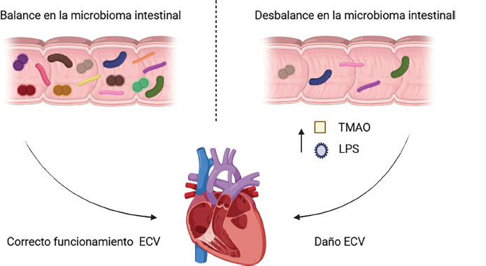
A balanced gut microbiota contributes to cardiovascular health. Conversely, dysbiosis generates substances such as trimethylamine N-oxide (TMAO) and lipopolysaccharides (LPS), which are associated with cardiovascular damage. This underscores the importance of maintaining microbial equilibrium to prevent cardiovascular diseases.
The interaction between the microbiota and the host is complex and dual in nature, where certain metabolic processes can promote endothelial health while others accelerate vascular damage. Table 2 summarizes this balance, contrasting the protective effects of short-chain fatty acids (SCFAs) and intestinal barrier integrity against the pathogenic effects derived from choline and carnitine metabolism, as well as systemic inflammation due to endotoxin translocation.
Table 2. Gut microbiota and cardiovascular disease: interplay between protective and pathogenic effects
Mechanism |
Protective Effects |
Pathogenic Effects |
References |
Dietary Fiber Metabolism |
Production of short-chain fatty acids (SCFAs: butyrate, propionate, acetate) reducing inflammation, improving blood pressure and insulin sensitivity. |
- |
|
Choline/Carnitine/Lecithin Metabolism |
- |
TMAO production promoting atherosclerosis, endothelial dysfunction, and thrombosis |
|
Gut Barrier Integrity |
Prevents endotoxin translocation and reduces systemic inflammation |
Dysbiosis increases intestinal permeability, allowing LPS entry and promoting chronic inflammation |
|
Immunomodulation |
Induction of T-regulatory cells and IL-10 production; reduced vascular inflammation |
Activation of proinflammatory cells (Th17, M1 macrophages) and elevation of proatherogenic cytokines |
|
Lipid metabolism |
Reduced serum cholesterol and bile acid modulation |
Alterations promoting atherogenic dyslipidemias |
|
Blood Pressure Regulation |
SCFAs activate vascular tone-regulating receptors (GPR41/GPR43) |
Dysbiosis associated with hypertension in animal models and humans |
|
Obesity/Metabolic Syndrome Impact |
Specific bacteria (Akkermansia muciniphila) improve metabolism and reduce adiposity |
Microbial imbalance linked to obesity, insulin resistance, and increased cardiovascular risk |
Discussion
A direct relationship exists between gut microbiota and cardiovascular diseases (CVD), presenting both opportunities and challenges for global public health. In middle- and low-income countries like Mexico, CVD accounts for 25% of annual deaths, compounded by risk factors such as obesity (39% in adults) and diabetes (14.4%), conditions strongly linked to gut dysbiosis78-79. Globally, CVD causes 18.6 million deaths annually, highlighting the urgent need for microbiota modulation as a cross-cutting strategy to address social and biological determinants72, 79.
Microbiota in the Latin American context
Accelerated shifts toward ultra-processed food consumption and persistent structural inequalities exacerbate cardiovascular risk across Latin America. Diets low in fiber and high in saturated fats induce dysbiosis, diminish short-chain fatty acid (SCFA) production, and promote proinflammatory microbial profiles associated with arterial stiffness and hypertension43, 51, 86. Regional dietary heterogeneity distinctly modulates microbiota composition: Central America relies on maize/beans (≥40% caloric intake), the Andes prioritize quinoa/tubers, and the Southern Cone exhibits high meat consumption (120 kg/capita/year). Notably, rural Guatemalan and Bolivian populations demonstrate 30% greater Bacteroidetes abundance and reduced TMAO levels linked to fiber-rich diets88.
In Mexico, only 43% of the population consumes fruits/vegetables daily, while 58% ingest sugar-sweetened beverages, disrupting Firmicutes/Bacteroidetes equilibrium78. These patterns necessitate evidence-based interventions. Promoting fiber-rich traditional foods (native maize, beans, amaranth) elevates Bacteroidetes and reduces TMAO by ≤40%, aligning with WHO recommendations (≥400g daily vegetables)71, 73, 80. This approach proves particularly relevant in rural Mexican communities with limited fresh food access, where nutrition education programs offer high cost-effectiveness76.
Regulating ultra-processed foods, comprising 70% of Mexico’s market, is equally critical. Chile’s front-of-package labeling (Law 20.606) reduced consumption by 24%, while urban agriculture initiatives in Medellín and Quito improved fresh produce access, providing transferable models for microbiota modulation84–85. Primary care innovations, such as targeted probiotics (Lactobacillus reuteri strains reducing LDL by 9% in high-risk groups), offer alternatives for marginalized populations with limited pharmaceutical access74.
Persistent structural barriers include: socioeconomic inequality (41.9% of Mexicans live in poverty, restricting prebiotic food access)81; a research gap (<5% of microbiota-CVD studies include Latin American cohorts)77; and insufficient healthcare infrastructure (80% of public hospitals lack specialized laboratories for advanced therapies like fecal microbiota transplantation)75, 82.
Global lessons inform actionable strategies: integrating TMAO biomarkers into clinical guidelines per American Heart Association recommendations could optimize secondary prevention, particularly for diabetic patients83. International collaborations with PAHO or the Global Heart Hub may facilitate funding and expertise. Multisectoral approaches, revitalizing ancestral fermented foods (e. g., Mexican pulque (fermented agave sap), tepache (fermented pineapple), through agricultural, educational, and economic policies, could bridge traditional practices with contemporary scientific evidence87.
Conclusion
The gut microbiota has emerged as a pivotal contributor to the pathophysiology of cardiovascular diseases (CVD). Its influence operates through complex mechanisms, including the production of proinflammatory metabolites such as trimethylamine N-oxide (TMAO), implicated in atherosclerosis progression, and the regulation of protective compounds like short-chain fatty acids (SCFAs), which modulate inflammation and enhance endothelial function.
Interventional strategies, including fiber-rich diets, targeted probiotics, and fecal microbiota transplantation (FMT), demonstrate potential to restore microbial homeostasis, attenuate inflammatory markers, improve lipid profiles, and ultimately reduce the global burden of CVD. Nevertheless, significant challenges persist: methodological heterogeneity across studies, the need to establish robust causal relationships, and barriers to implementing sustainable, equitable public health strategies.
Future advancements necessitate integrating gut microbiota into cardiovascular risk prediction models and developing personalized nutritional, pharmacological, and microbiological interventions. Research must prioritize robust clinical trials and public policies that translate scientific insights into actionable solutions. Within this paradigm, the gut microbiota represents not only a critical systemic modulator but also an innovative avenue for addressing one of the world’s leading causes of mortality.
Acknowledgments
A. A. G. M. gratefully acknowledges the academic support provided by the Doctoral Program in Biomedical and Molecular Biotechnology Sciences at the Escuela Nacional de Ciencias Biológicas (ENCB) of the Instituto Politécnico Nacional (IPN), which facilitated the completion of this work. Special recognition is extended to the Secretaría de Ciencia, Humanidades, Tecnología e Innovación for the postgraduate scholarship (CVU 1276564).
Authors’ contributions
Conceptualization and study design: M. A. S. G. and A. A. G. M. Drafting the initial manuscript: M. A. S. G. and A. A. G. M. All authors reviewed the results and approved the final version of the manuscript.
Funding
This research did not receive any specific grant from funding agencies in the public, commercial, or not-for-profit sectors.
Competing interests
The authors declare no competing interests
Disclaimer / Editor's Note
The ideas, opinions, and data expressed in published articles are the sole responsibility of their authors and contributors and do not necessarily represent the position of Ciencia y Salud, the editorial team, or the Instituto Tecnológico de Santo Domingo (INTEC). Ciencia y Salud, its editors, and INTEC assume no responsibility for any damage to persons or property that may result from the use of the methods, procedures, instructions, or products mentioned in the published content.
References
1. Brown JM, Hazen SL. Microbial modulation of cardiovascular disease. Nat Rev Microbiol. 2018;16(3):171-81. https://doi.org/10.1038/nrmicro.2017.149.
2. Jie Z, Xia H, Zhong SL, Feng Q, Li S, Liang S, et al. The gut microbiome in atherosclerotic cardiovascular disease. Nat Commun. 2017;8:845. https://doi.org/10.1038/s41467-017-00900-1
3. Tang WHW, Li DY, Hazen SL. Dietary metabolism, gut microbiota, and acute heart failure. Heart Fail Rev. 2017;22(2):217-24. https://doi.org/10.1038/s41569-018-0108-7
4. Tang WHW, Bäckhed F, Landmesser U, Hazen SL. Intestinal microbiota in cardiovascular health and disease: JACC state-of-the-art review. J Am Coll Cardiol. 2019;73(16):2089-105. https://doi.org/10.1016/j.jacc.2019.03.024
5. Witkowski M, Weeks TL, Hazen SL. Gut microbiota and cardiovascular disease. Circ Res. 2020;127(4):553-70. https://doi.org/10.1161/CIRCRESAHA.120.316242
6. World Health Organization. Cardiovascular diseases (CVDs) [Internet]. Geneva: WHO; 2021. [citado 2025 Ene 15]. Available from: https://www.who.int/news-room/fact-sheets/detail/cardiovascular-diseases-(cvds).
7. Blaser MJ. Antibiotic use and its consequences for the normal microbiome. Science. 2016;352(6285):544-5. https://doi.org/10.1126/science.aad9358
8. Rodrigues Hoffmann A, Proctor LM, Surette MG, Suchodolski JS. The microbiome: The trillions of microorganisms that maintain health and cause disease in humans and companion animals. Vet Pathol. 2016;53(1):10-21. https://doi.org/10.1177/0300985815595517
9. Wang L, Wang S, Zhang Q, He C, Fu C, Wei Q. The role of the gut microbiota in health and cardiovascular diseases. Mol Biomed. 2022;3(1):30. https://doi.org/10.1186/s43556-022-00091-2
10. Valdes AM, Walter J, Segal E, Spector TD. Role of the gut microbiota in nutrition and health. BMJ. 2018;361: k2179. https://doi.org/10.1136/bmj.k2179.
11. Cryan JF, O'Riordan KJ, Sandhu K, Peterson V, Dinan TG. The gut microbiome in neurological disorders. Lancet Neurol. 2020;19(2):179-94. https://doi.org/10.1016/S1474-4422(19)30356-4
12. Sender R, Fuchs S, Milo R. Revised estimates for the number of human and bacteria cells in the body. PLoS Biol. 2016 Aug;14(8): e1002533. https://doi.org/10.1371/journal.pbio.1002533
13. Magne F, Gotteland M, Gauthier L, Zazueta A, Pesoa S, Navarrete P, et al. The Firmicutes/Bacteroidetes ratio: A relevant marker of gut dysbiosis in obese patients. Nutrients. 2020 May;12(5):1474. https://doi.org/10.3390/nu12051474
14. Miele L, Giorgio V, Alberelli MA, De Candia E, Gasbarrini A, Grieco A. Impact of gut microbiota on obesity, diabetes, and cardiovascular disease risk. Curr Cardiol Rep. 2015 Dec;17(12):120. https://doi.org/10.1007/s11886-015-0671-z
15. Sonnenburg, J., Bäckhed, F. Diet–microbiota interactions as moderators of human metabolism. Nature 535, 56–64 (2016). https://doi.org/10.1038/nature18846
16. Sonnenburg ED, Sonnenburg JL. The ancestral and industrialized gut microbiota and implications for human health. Nat Rev Microbiol. 2019 Jun;17(6):383-90. https://doi.org/10.1038/s41579-019-0191-8
17. Thaiss CA, Zmora N, Levy M, Elinav E. The microbiome and innate immunity. Nature. 2016 Jul;535(7610):65-74. https://doi.org/10.1038/nature18847
18. De Filippo C, Cavalieri D, Di Paola M, Ramazzotti M, Poullet JB, Massart S, et al. Impact of diet in shaping gut microbiota revealed by a comparative study in children from Europe and rural Africa. Proc Natl Acad Sci U S A. 2010 Aug;107(33):14691-6. https://doi.org/10.1073/pnas.1005963107
19. Floch MH, Ringel Y, Walker WA, editors. The microbiota in gastrointestinal pathophysiology. London: Elsevier; 2016. p. 227-32. ISBN: 9780128040621.
20. Azzini E, Polito A, Fumagalli A, Intorre F, Venneria E, Durazzo A, et al. Mediterranean diet effect: An Italian picture. Nutr J. 2011;10:125. https://doi.org/10.1186/1475-2891-10-125
21. Chassaing B, Koren O, Goodrich JK, Poole AC, Srinivasan S, Ley RE, et al. Dietary emulsifiers impact the mouse gut microbiota promoting colitis and metabolic syndrome. Nature. 2015 Mar;519(7541):92-6. https://doi.org/10.1038/nature14232
22. Suez J, Korem T, Zeevi D, Zilberman-Schapira G, Thaiss CA, Maza O, et al. Artificial sweeteners induce glucose intolerance by altering the gut microbiota. Nature. 2014 Oct;514(7521):181-6. https://doi.org/10.1038/nature13793
23. Palmer C, Bik EM, DiGiulio DB, Relman DA, Brown PO. Development of the human infant intestinal microbiota. PLoS Biol. 2007 Jul;5(7): e177. https://doi.org/10.1371/journal.pbio.0050177
24. Jakobsson HE, Jernberg C, Andersson AF, Sjölund-Karlsson M, Jansson JK, Engstrand L. Short-term antibiotic treatment has differing long-term impacts on the human throat and gut microbiome. PLoS One. 2010;5(3): e9836. https://doi.org/10.1371/journal.pone.0009836
25. Blaser MJ. Stop the killing of beneficial bacteria. Nature. 2011 Aug;476(7361):393-4. https://doi.org/10.1038/476393a
26. Kundu P, Blacher E, Elinav E, Pettersson S. Our gut microbiome: The evolving inner self. Cell. 2017 Dec;171(7):1481-93. https://doi.org/10.1016/j.cell.2017.11.024
27. Claesson MJ, Cusack S, O'Sullivan O, Greene-Diniz R, de Weerd H, Flannery E, et al. Composition, variability, and temporal stability of the intestinal microbiota of the elderly. Proc Natl Acad Sci U S A. 2011;108 Suppl 1:4586-91. https://doi.org/10.1073/pnas.1000097107
28. Langille MGI, Meehan CJ, Koenig JE, Dhanani AS, Rose RA, Howlett SE, et al. Microbial shifts in the aging mouse gut. Microbiome. 2014;2(1):50. https://doi.org/10.1186/s40168-014-0050-9
29. Claesson MJ, Jeffery IB, Conde S, Power SE, O’Connor EM, Cusack S, et al. Gut microbiota composition correlates with diet and health in the elderly. Nature. 2012 Aug;488(7410):178-84. https://doi.org/10.1038/nature11319
30. Biagi E, Franceschi C, Rampelli S, Severgnini M, Ostan R, Turroni S, et al. Gut microbiota and extreme longevity. Curr Biol. 2016 Jun;26(11):1480-5. https://doi.org/10.1016/j.cub.2016.04.016
31. Moya A, Ferrer M. Functional redundancy-induced stability of gut microbiota subjected to disturbance. Trends Microbiol. 2016 May;24(5):402-13. https://doi.org/10.1016/j.tim.2016.02.002
32. Faith JJ, Guruge JL, Charbonneau M, Subramanian S, Seedorf H, Goodman AL, et al. The long-term stability of the human gut microbiota. Science. 2013;341(6141):1237439. https://doi.org/10.1126/science.1237439
33. Buttó LF, Schaubeck M, Haller D. Mechanisms of microbe-host interaction in Crohn’s disease: Dysbiosis vs. pathobiont selection. Front Immunol. 2015;6:555. https://doi.org/10.3389/fimmu.2015.00555
34. Gevers D, Kugathasan S, Denson LA, Vazquez-Baeza Y, Van Treuren W, Ren B, et al. The treatment-naive microbiome in new-onset Crohn's disease. Cell Host Microbe. 2014 Mar;15(3):382-92. https://doi.org/10.1016/j.chom.2014.02.005
35. Jostins L, Ripke S, Weersma RK, Duerr RH, McGovern DP, Hui KY, et al. Host-microbe interactions have shaped the genetic architecture of inflammatory bowel disease. Nature. 2012 Nov;491(7422):119-24. https://doi.org/10.1038/nature11582
36. Schaubeck M, Clavel T, Calasan J, Lagkouvardos I, Haange SB, Jehmlich N, et al. Dysbiotic gut microbiota causes transmissible Crohn's disease-like ileitis independent of failure in antimicrobial defence. Gut. 2016;65(2):225-37. https://doi.org/10.1136/gutjnl-2015-309333
37. Kamada N, Chen GY, Inohara N, Núñez G. Control of pathogens and pathobionts by the gut microbiota. Nat Immunol. 2013;14(7):685-90. https://doi.org/10.1038/ni.2608
38. Druml W. Intestinal cross-talk: The gut as motor of multiple organ failure. Med Klin Intensivmed Notfmed. 2018;113(6):470-7. https://doi.org/10.1007/s00063-018-0475-1
39. Andersen K, Kesper MS, Marschner JA, Konrad L, Ryu M, Kumar Vr S, et al. Intestinal dysbiosis, barrier dysfunction, and bacterial translocation account for CKD-related systemic inflammation. J Am Soc Nephrol. 2017;28(1):76-83. https://doi.org/10.1681/ASN.2015111285
40. Santisteban MM, Qi Y, Zubcevic J, Kim S, Yang T, Shenoy V, et al. Hypertension-linked pathophysiological alterations in the gut. Circ Res. 2017;120(2):312-23. https://doi.org/10.1161/CIRCRESAHA.116.309006
41. Zhou X, Li J, Guo J, Geng B, Ji W, Zhao Q, et al. Gut-dependent microbial translocation induces inflammation and cardiovascular events after ST-elevation myocardial infarction. Microbiome. 2018;6(1):66. https://doi.org/10.1186/s40168-018-0441-4
42. Li XS, Obeid S, Klingenberg R, Gencer B, Mach F, Räber L, et al. Gut microbiota-dependent trimethylamine N-oxide in acute coronary syndromes: A prognostic marker for incident cardiovascular events beyond traditional risk factors. Eur Heart J. 2017;38(11):814-24. https://doi.org/10.1093/eurheartj/ehw582
43. Koeth RA, Wang Z, Levison BS, Buffa JA, Org E, Sheehy BT, et al. Intestinal microbiota metabolism of L-carnitine, a nutrient in red meat, promotes atherosclerosis. Nat Med. 2013;19(5):576-85. https://doi.org/10.1038/nm.3145
44. Wang B, Qiu J, Lian J, Yang X, Zhou J. Gut metabolite trimethylamine-N-oxide in atherosclerosis: From mechanism to therapy. Front Cardiovasc Med. 2021;8:723886. https://doi.org/10.3389/fcvm.2021.723886
45. Li X, Liu Y, Yang X, Li C, Song Z. The oral microbiota: Community composition, influencing factors, pathogenesis, and interventions. Front Microbiol. 2022;13:895537. https://doi.org/10.3389/fmicb.2022.895537
46. Heianza Y, Ma W, Manson JE, Rexrode KM, Qi L. Gut Microbiota Metabolites and Risk of Major Adverse Cardiovascular Disease Events and Death: A Systematic Review and Meta-Analysis of Prospective Studies. Circulation. 2021;143(12):1204-12. https://doi.org/10.1161/JAHA.116.004947
47. Wang Z, Roberts AB, Buffa JA, Levison BS, Zhu W, Org E, et al. Non-lethal inhibition of gut microbial trimethylamine production for the treatment of atherosclerosis. J Am Coll Cardiol. 2021;77(18):2300-12. Https://doi.org/10.1016/j.cell.2015.11.055
48. Dinser L, Meisinger C, Amann U, Heier M, Thilo C, Kuch B, et al. Peripheral arterial disease is associated with higher mortality in patients with incident acute myocardial infarction. Eur J Intern Med. 2018;51:46-52. https://doi.org/10.1016/j.ejim.2018.01.007
49. Silva YP, Bernardi A, Frozza RL. The role of short-chain fatty acids from gut microbiota in gut-brain communication. Front Endocrinol. 2020;11:25. https://doi.org/10.3389/fendo.2020.00025
50. Marques FZ, Nelson E, Chu PY, Horlock D, Fiedler A, Ziemann M, et al. High-fiber diet and acetate supplementation change the gut microbiota and prevent the development of hypertension and heart failure in hypertensive mice. Hypertension. 2017;70(3):479-85. https://doi.org/10.1161/HYPERTENSIONAHA.117.08909
51. Pluznick JL, Protzko RJ, Gevorgyan H, Peterlin Z, Sipos A, Han J, et al. Olfactory receptor responding to gut microbiota-derived signals plays a role in renin secretion and blood pressure regulation. Proc Natl Acad Sci U S A. 2013;110(11):4410-5. https://doi.org/10.1073/pnas.1215927110
52. Maruvada P, Leone V, Kaplan LM, Chang EB. The human microbiome and obesity: Moving beyond associations. Cell Host Microbe. 2017;22(5):589-99. https://doi.org/10.1016/j.chom.2017.10.005
53. Sun J, Yuan B, Wu Y, Gong Y, Guo W, Fu S, et al. Sodium butyrate protects N2a cells against Aβ toxicity in vitro. Mediators Inflamm. 2020;2020:7605160. https://doi.org/10.1155/2020/7605160
54. Natarajan A, Zlitni S, Brooks EF, Vance SE, Dahlen A, Hedlin H, et al. Gastrointestinal symptoms and fecal shedding of SARS-CoV-2 RNA suggest prolonged gastrointestinal infection. Med. 2022;3(6):371-87. https://doi.org/10.1016/j.medj.2022.04.001
55. Pasini E, Aquilani R, Testa C, Baiardi P, Angioletti S, Boschi F, et al. Pathogenic gut flora in patients with chronic heart failure. J Clin Med. 2020;9(3):1-15. https://doi.org/10.1016/j.jchf.2015.10.009
56. Witkowski M, Weeks TL, Hazen SL. Gut microbiota and cardiovascular disease. Science. 2020;368(6491):577-82. https://doi.org/10.1161/CIRCRESAHA.120.316242
57. Prins FM, Collij V, Groot HE, Björk JR, Swarte JC, Andreu-Sánchez S, et al. The gut microbiome across the cardiovascular risk spectrum. Eur J Prev Cardiol. 2024;31(8):935-44. https://doi.org/10.1093/eurjpc/zwad377
58. Lu Q, Wang Y, Li X, Zhang Y, Zhang H, Wang X, et al. Gut microbiota-derived secondary bile acids, bile acids receptor polymorphisms, and risk of cardiovascular disease in individuals with newly diagnosed type 2 diabetes: A cohort study. Am J Clin Nutr. 2024;119(2):324-32. https://doi.org/10.3389/fimmu.2023.1127743
59. Koh A, De Vadder F, Kovatcheva-Datchary P, Bäckhed F. From dietary fiber to host physiology: short-chain fatty acids as key bacterial metabolites. Cell. 2016;165(6):1332-45. https://doi.org/10.1016/j.cell.2016.05.041
60. Tang WHW, Kitai T, Hazen SL. Gut microbiota in cardiovascular health and disease. Circ Res. 2017;120(7):1183-96. https://doi.org/10.1161/CIRCRESAHA.117.309715
61. Wang Z, Klipfell E, Bennett BJ, Koeth R, Levison BS, Dugar B, et al. Gut flora metabolism of phosphatidylcholine promotes cardiovascular disease. Nature. 2011;472(7341):57-63. https://doi.org/10.1038/nature09922
62. Cani PD, Amar J, Iglesias MA, Poggi M, Knauf C, Bastelica D, et al. Metabolic endotoxemia initiates obesity and insulin resistance. Diabetes. 2007;56(7):1761-72. https://doi.org/10.2337/db06-1491
63. Leclercq S, Matamoros S, Cani PD, Neyrinck AM, Jamar F, Stärkel P, et al. Intestinal permeability, gut-bacterial dysbiosis, and behavioral markers of alcohol-dependence severity. Proc Natl Acad Sci USA. 2014;111(42):E4485-93. https://doi.org/10.1073/pnas.1415174111
64. Li X, Deroide N, Mallat Z. The role of the inflammasome in cardiovascular diseases. J Mol Med. 2014;92(4):307-19. https://doi.org/10.1007/s00109-014-1144-3
65. Miura K, Ohnishi H. Role of gut microbiota and Toll-like receptors in nonalcoholic fatty liver disease. World J Gastroenterol. 2014;20(23):7381-91. https://doi.org/10.3748/wjg.v20.i23.7381
66. Gérard P. Gut microbiota and obesity. Cell Mol Life Sci. 2016;73(1):147- https://doi.org/10.1007/s00018-015-2061-5
67. Velagapudi VR, Hezaveh R, Reigstad CS, Gopalacharyulu P, Yetukuri L, Islam S, et al. The gut microbiota modulates host energy and lipid metabolism in mice. J Lipid Res. 2010;51(5):1101-12. https://doi.org/10.1194/jlr.M002774
68. Muralitharan RR, Jama HA, Xie L, Peh A, Snelson M, Marques FZ. Microbial Peer Pressure: The Role of the Gut Microbiota in Hypertension and Its Complications. Hypertension. 2020;76(6):1674 87. https://doi.org/10.1161/HYPERTENSIONAHA.120.14473
69. Dao MC, Everard A, Aron-Wisnewsky J, Sokolovska N, Prifti E, Verger EO, et al. Akkermansia muciniphila and improved metabolic health during a dietary intervention in obesity: relationship with gut microbiome richness and ecology. Gut. 2016;65(3):426-36. https://doi.org/10.1136/gutjnl-2014-308778
70. Turnbaugh PJ, Ley RE, Mahowald MA, Magrini V, Mardis ER, Gordon JI. An obesity-associated gut microbiome with increased capacity for energy harvest. Nature. 2006;444(7122):1027-31. https://doi.org/10.1038/nature05414
71. Janeiro MH, Ramírez MJ, Milagro FI, Martínez JA, Solas M. Implication of trimethylamine N-oxide (TMAO) in disease: Potential biomarker or new therapeutic target. Nutrients. 2018;10(10):1398. https://doi.org/10.3390/nu10101398
72. Ahmad AF, Dwivedi G, O'Gara F, Caparros-Martin J, Ward NC. The gut microbiome and cardiovascular disease: current knowledge and clinical potential. Am J Physiol Heart Circ Physiol. 2019 Nov 1;317(5): H923-H938. https://doi.org/10.1152/ajpheart.00376.2019
73. De Filippis F, Pellegrini N, Vannini L, Jeffery IB, La Storia A, Laghi L, Serrazanetti DI, Di Cagno R, Ferrocino I, Lazzi C, Turroni S, Cocolin L, Brigidi P, Neviani E, Gobbetti M, O'Toole PW, Ercolini D. High-level adherence to a Mediterranean diet beneficially impacts the gut microbiota and associated metabolome. Gut. 2016 Nov; 65(11):1812-1821. https://doi.org/10.1136/gutjnl-2015-309957
74. Khalesi S, Bellissimo N, Vandelanotte C, Williams S, Stanley D, Irwin C. A review of probiotic supplementation in healthy adults: Helpful or hype? Eur J Clin Nutr. 2019;73(1):24-37. https://doi.org/10.1038/s41430-018-0135-9
75. Vrieze A, Van Nood E, Holleman F, Salojärvi J, Kootte RS, Bartelsman JF, Dallinga-Thie GM, Ackermans MT, Serlie MJ, Oozeer R, Derrien M, Druesne A, Van Hylckama Vlieg JE, Bloks VW, Groen AK, Heilig HG, Zoetendal EG, Stroes ES, de Vos WM, Hoekstra JB, Nieuwdorp M. Transfer of intestinal microbiota from lean donors increases insulin sensitivity in individuals with metabolic syndrome. Gastroenterology. 2012 Oct;143(4):913-6. e7. https://doi.org/10.1053/j.gastro.2012.06.031
76. World Health Organization. Healthy diet [Internet]. Geneva: WHO; 2020 – Disponible en: https://www.who.int/news-room/fact-sheets/detail/healthy-diet
77. Valdes AM, Walter J, Segal E, Spector TD. Role of the gut microbiota in nutrition and health. BMJ. 2018;361: k2179. https://doi.org/10.1136/bmj.k2179.
78. Instituto Nacional de Salud Pública. Encuesta Nacional de Salud y Nutrición (ENSANUT 2021) [Internet]. México: INSP; 2021 [citado 2025 Ene 10]. Disponible en: https://ensanut.insp.mx/
79. Instituto Nacional de Estadística y Geografía. Estadísticas de mortalidad en México [Internet]. México: INEGI; 2023 [citado 2024 Ene 25]. Disponible en: https://www.inegi.org.mx/
80. Organización Mundial de la Salud. Enfermedades cardiovasculares: Datos y cifras [Internet]. 2021. Disponible en: https://www.who.int/es/
81. Consejo Nacional de Evaluación de la Política de Desarrollo Social. Medición de la pobreza en México 2022: Resultados nacionales [Internet]. México: CONEVAL; 2022 [citado 2025 Ene 29]. Disponible en: https://www.coneval.org.mx/Medicion/Paginas/PobrezaInicio.aspx
82. Secretaría de Salud. Informe anual del sistema de salud público mexicano [Internet]. [citado 2025 Ene 29]. Disponible en: https://www.gob.mx/salud/.
83. Tang WHW, Li DY, Hazen SL. Dietary metabolism, the gut microbiome, and heart failure. Nat Rev Cardiol. 2019 Mar;16(3):137-154. https://doi.org/10.1038/s41569-018-0108-7
84. Instituto Nacional de Salud Pública. Consumo de alimentos ultraprocesados y su asociación con obesidad en México: Reporte técnico 2022 [Internet]. [citado 2025 Feb 28]. Disponible en: https://www.insp.mx/produccion-editorial/nuevas-publicaciones/consumo-alimentos-ultraprocesados
85. Ministerio de Salud de Chile. Informe de evaluación de la implementación de la Ley de Etiquetado de Alimentos (Ley 20.606) 2021 [Internet]. [citado 2025 Feb 18] Disponible en: https://www.minsal.cl/wp-content/uploads/2021/05/Informe-Evaluacion-Ley-20606-2021.pdf
86. Sonnenburg ED, Sonnenburg JL. Starving our microbial self: the deleterious consequences of a diet deficient in microbiota-accessible carbohydrates. Cell Metab. 2014;20(5):779-86. https://doi.org/10.1016/j.cmet.2014.07.003
87. Perrone P, D'Angelo S. Gut microbiota modulation through Mediterranean diet foods: implications for human health. Nutrients. 2025;17(6):948. https://doi.org/10.3390/nu17060948
88. Martínez I, Stegen JC, Maldonado-Gómez MX, Eren AM, Siba PM, Greenhill AR, et al. The gut microbiota of rural Papua New Guineans: composition, diversity patterns, and ecological processes. Cell Rep. 2015 Apr 28;11(4):527-38. https://doi.org/10.1016/j.celrep.2015.03.049
_______________________________
1 Research and Education Department, Universidad del Valle de Puebla, México. ORCID: https://orcid.org/0000-0003-0902-8993, email: mildred.sauce@uvp.edu.mx
2 Experimental Pathology Research Laboratory, Hospital Infantil de México Federico Gómez; Clinical and Environmental Research Laboratory; Escuela Nacional de Ciencias Biológicas, IPN, México. ORCID: https://orcid.org/0000-0001-5172-703X, email: aguerram2300@alumno.ipn.mx