Ciencia y Salud, Vol. 9, No. 3, septiembre-diciembre, 2025 • ISSN (impreso): 2613-8816 • ISSN (en línea): 2613-8824
CLINICAL PRESENTATION AND MANAGEMENT OF PHACE SYNDROME IN A PEDIATRIC PATIENT: A CASE REPORT. PHACE SYNDROME IN PEDIATRICS
Presentación clínica y manejo del síndrome PHACE en un paciente pediátrico: Reporte de un Caso. Síndrome PHACE en pediatría
DOI: https://doi.org/10.22206/cisa.2025.v9i3.3499
Enviado: 21 de marzo 2025 ● Aceptado: 23 de abril 2025

Cómo citar: Capestany, C., Báez, K., Vásquez, L., Ortiz Hernández, I. (2025). Clinical Presentation and Management of PHACE Syndrome in a Pediatric patient: A Case Report. PHACE Syndrome in Pediatrics. Ciencia y Salud, 9(3), 99-105. https://doi.org/10.22206/cisa.2025.v9i3.3499
Abstract
Introduction. PHACE syndrome is a rare neurocutaneous disorder characterized by posterior fossa malformations, hemangiomas, arterial anomalies, cardiac defects, and ocular abnormalities. Propranolol is an effective treatment for hemangiomas, but cerebrovascular and cardiac assessments are recommended before initiating therapy. Long-term treatment is often necessary, with a risk of rebound growth upon discontinuation. Case Description. We present the case of a 4-year-old girl, born at 41 weeks, with prenatal exposure to alcohol and marijuana. Initially normal, she developed facial hemangiomas at 20 days, leading to eye obstruction and lip ulceration. Subsequently, imaging revealed mega cisterna magna, periorbital changes, corpus callosum dysgenesis, and possible Dandy-Walker malformation. At 12 months, she was diagnosed with PHACE syndrome based on hemangioma size, posterior fossa anomalies, and ocular findings. By 20 months, she exhibited delayed motor skills, hypotonia, and convergent strabismus. At 4 years, she presents severe developmental delays. Propranolol effectively reduced her hemangiomas, with dosage adjustments based on weight changes. She receives multidisciplinary care, although financial constraints limit access to imaging and laboratory tests. Discussion. PHACE syndrome primarily affects females, with around 400 documented cases globally. Facial hemangiomas are present in 20-30% of these cases. Diagnosis is based on clinical evaluation and imaging studies, including MRI and echocardiography. While propranolol is the preferred treatment, long-term outcomes remain uncertain, highlighting the need for ongoing monitoring of cerebrovascular anomalies. Conclusion. This case underscores the complexity of PHACE syndrome, highlighting the need for early diagnosis and careful management. Further research is required to improve treatment options.
Keywords: Neurocutaneous disorder, propanolol, PHACE syndrome.
Resumen
Introducción. El síndrome PHACE es un raro trastorno neurocutáneo que se caracteriza por malformaciones de la fosa posterior, hemangiomas, anomalías arteriales, defectos cardíacos y anomalías oculares. El propranolol es un tratamiento eficaz para los hemangiomas, pero se recomienda realizar evaluaciones cerebrovasculares y cardíacas antes de iniciar la terapia. A menudo, se requiere un tratamiento a largo plazo, con riesgo de crecimiento rebote al interrumpirlo. Descripción del caso. Presentamos el caso de una niña de 4 años, nacida a las 41 semanas, con exposición prenatal a alcohol y marihuana. Inicialmente normal, desarrolló hemangiomas faciales a los 20 días, lo que provocó obstrucción ocular y ulceración labial. Posteriormente, las imágenes revelaron mega cisterna magna, cambios periorbitales, disgenesia del cuerpo calloso y posible malformación de Dandy-Walker. A los 12 meses, se diagnosticó síndrome PHACE, basado en el tamaño del hemangioma, anomalías de la fosa posterior y hallazgos oculares. A los 20 meses, mostró retraso en habilidades motoras, hipotonía y estrabismo convergente. A los 4 años, presenta retrasos severos en el desarrollo. El propranolol redujo eficazmente sus hemangiomas, con ajustes de dosis según cambios de peso. Recibe atención multidisciplinaria, aunque las limitaciones financieras restringen el acceso a imágenes y pruebas de laboratorio. Discusión. El síndrome PHACE afecta principalmente a mujeres, con aproximadamente 400 casos documentados a mundialmente. Los hemangiomas faciales están presentes en el 20-30% de estos casos. El diagnóstico se basa en la evaluación clínica y estudios de imagen, incluyendo resonancia magnética y ecocardiografía. Aunque el propranolol es el tratamiento preferido, los resultados a largo plazo siguen siendo inciertos, lo que resalta la necesidad de un monitoreo continuo de las anomalías cerebrovasculares. Conclusión. Este caso destaca la complejidad del síndrome PHACE, enfatizando la necesidad de un diagnóstico temprano y una gestión cuidadosa. Se requiere más investigación para mejorar las opciones de tratamiento.
Palabras clave: Propanolol, síndrome de PHACE, trastorno neurocutáneo.
Introduction
Infantile hemangiomas (IH) are the most common benign vascular tumors in infancy, typically presenting during the first few weeks of life. Large, segmental IHs may serve as cutaneous markers for underlying structural anomalies in other organ systems. PHACE syndrome, a neurocutaneous disorder first described by Frieden et al. in 1996, is characterized by the association of segmental IHs with a spectrum of cerebrovascular, cardiovascular, and ocular abnormalities. The syndrome’s diagnostic criteria were updated in 2016, introducing major and minor criteria to account for its clinical heterogeneity1, 2.
Oral propranolol has significantly transformed the management of IH, yet its use in patients with PHACE syndrome requires careful consideration due to potential risks such as exacerbation of vascular anomalies and complications associated with cerebrovascular insufficiency. Patients with PHACE syndrome often necessitate extended propranolol therapy and face a heightened risk of rebound growth after treatment cessation. While advancements in the understanding of IH pathophysiology and propranolol mechanisms have improved clinical outcomes, the genetic underpinnings and exact pathogenesis of PHACE syndrome remain incompletely elucidated3, 4.
This case report details a patient with PHACE syndrome, highlighting diagnostic challenges, therapeutic strategies, and long-term management considerations. The discussion underscores the importance of a multidisciplinary approach to optimize outcomes for this rare and complex condition.
Case description
We present the case of a 4-year-old Dominican girl born at 41 weeks’ gestation via cesarean section, with a clinical history notable for multiple congenital anomalies. Maternal history revealed alcohol consumption during the first trimester, as well as exposure to marijuana and electronic cigarette smoke. Prenatal care was delayed due to the COVID-19 pandemic.
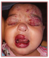
At birth, the patient exhibited erythema on both cheeks, right eyelid ptosis, and clinical suspicion of hydrocephalus (Figures 1 and 2). A cranial computed tomography (CT) scan identified a mega cisterna magna and periorbital soft tissue abnormalities. A follow-up CT revealed additional findings, including dysgenesis of the corpus callosum, possible Dandy-Walker malformation, enlarged lacrimal glands, and asymmetry of the parotid glands, with hypertrophy of the right parotid gland. Magnetic resonance imaging (MRI) confirmed hydrocephalus and posterior fossa anomalies consistent with Dandy-Walker malformation. Hydrocephalus was surgically managed with the placement of a ventriculoperitoneal shunt at 7 months of age.
At 12 months, the patient was referred to a pediatric geneticist for evaluation of a suspected syndromic etiology. Physical examination revealed small, asymmetric eyes with limited voluntary opening, convergent strabismus, hyperemic conjunctiva, asymmetric lips, hemangiomas involving the upper and lower lips, a large irregular hemangioma spanning the neck and frontal scalp, isolated vascular papules in the ocular and labial regions, and hemangiomas on the eyelids. Based on clinical findings, a diagnosis of PHACE syndrome was established, supported by the presence of a segmental facial hemangioma >5 cm in diameter, posterior fossa anomalies, and ocular abnormalities.
Figure 1. Clinical presentation of the patient at one month of age
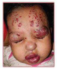
Figure 2. Clinical presentation of the patient at one month of age
Propranolol therapy was initiated at a dose of 10 mg, diluted in water and administered orally twice daily. Within two weeks of treatment, notable regression of hemangiomas was observed, particularly in the frontotemporal region, which significantly improved eye opening.
At 20 months, the patient demonstrated delayed motor milestones, stereotyped upper limb and head movements, marked hypotonia, and persistent convergent strabismus. Neurological and ophthalmological evaluations were conducted. Ocular ultrasonography identified posterior vitreous detachment and mild optic nerve excavation bilaterally. The patient was prescribed nutritional supplementation (L-carnitine, iron, folic acid, and vitamins A, C, and B-complex) alongside recommendations for early stimulation and physical therapy at home. Ophthalmological management included corrective lenses for strabismus and lubricating eye drops.
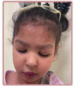
At 4 years of age, the patient presents with profound developmental delays, characterized by the inability to ambulate or communicate verbally. Despite these challenges, continued propranolol therapy has achieved substantial reduction in cutaneous hemangiomas (Figures 3 and 4). The patient remains under multidisciplinary follow-up, including neurosurgery, cardiology, genetics, and ophthalmology. Cardiologic evaluations, including echocardiography and electrocardiogram (ECG), have been unremarkable, allowing for safe continuation of propranolol therapy with weight-based dose adjustments. Current interventions include occupational therapy, speech therapy, and the use of orthopedic boots for support.
Figure 3. Clinical presentation of the patient at four years of age
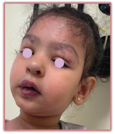
Figure 4. Clinical presentation of the patient at four years of age
Discussion
Infantile hemangiomas (IH) typically progress through three well-defined stages: proliferation, stabilization, and spontaneous involution. Hemangiomas located in specific facial regions are associated with an increased risk of ocular or central nervous system (CNS) involvement, potentially heightening the risk of cerebrovascular complications, such as stroke. Haggström et al. classified the face into four anatomic segments, frontotemporal (S1), maxillary (S2), mandibular (S3), and frontonasal (S4), to correlate hemangioma location with systemic anomalies. Segmental hemangiomas in S1 and S4 demonstrate a stronger association with cerebrovascular, cerebral, or ocular anomalies, whereas S3 hemangiomas are more commonly linked to cardiac anomalies, aortic arch malformations, and ventral developmental defects5-7.
PHACE syndrome is a rare neurocutaneous vascular condition that occurs in approximately 31% of patients with segmental facial hemangiomas larger than 5 cm. The syndrome has been documented in approximately 400 cases worldwide, predominantly in females, with a female-to-male ratio of 9:1. Despite increasing recognition, the true incidence remains unclear due to underreporting. The clinical and phenotypic heterogeneity of PHACE syndrome continues to present diagnostic and management challenges. While its etiology remains elusive, ongoing genetic investigations, such as the Stanford University study initiated in 2009 and projected to continue through 2030, aim to elucidate its molecular underpinnings5, 6.
Structural and vascular anomalies, particularly involving the CNS, are frequently reported in PHACE syndrome and emphasize the importance of multidisciplinary evaluations. Cranial imaging modalities, including MRI, gadolinium-enhanced MR angiography, and echocardiography, are essential for identifying associated malformations, such as Dandy-Walker malformation, cortical dysplasia, agenesis of the corpus callosum, and arterial anomalies, which predispose patients to stroke. CNS findings in PHACE patients often necessitate tailored interventions based on functional impact. Additionally, dental root anomalies, enamel hypoplasia, and lip hemangiomas, including ulcerated lesions, are not uncommon and require specialized dental and dermatological management. Ulcerated or non-ulcerated lip hemangiomas are rarely reported but carry a high risk of late-onset recurrent growth and ulceration post-treatment6, 7.
Propranolol, introduced as a transformative treatment for IH in 2008, has become the cornerstone of therapy for segmental hemangiomas, including those associated with PHACE syndrome. Its efficacy in reducing hemangioma size and halting progression is well-documented, and it has largely supplanted corticosteroids in this context. However, the initial adoption of propranolol in PHACE syndrome was cautious, given concerns about exacerbating cerebrovascular anomalies and stroke risk. Subsequent studies have demonstrated its safety profile, including minimal effects on blood pressure and perfusion in infants. A 2019 cohort study involving 76 PHACE patients treated with propranolol reported no instances of stroke, underscoring the importance of pre-treatment screening with echocardiography and physical examinations to rule out contraindications such as aortic arch coarctation4, 8.
Propranolol therapy in PHACE syndrome should be initiated with caution, starting at a low dose of 0.5–1 mg/kg/day, divided into three daily doses, and titrated to a therapeutic range of 2–3 mg/kg/day over the first week. Gradual dose escalation minimizes the risk of hemodynamic instability and potential cerebrovascular events. Treatment duration should be individualized, with close monitoring to assess for rebound growth, a well-documented phenomenon following premature discontinuation. Furthermore, neurosurgical options are considered for cerebrovascular disease9.
Recent research has explored the long-term outcomes of PHACE syndrome. A multicenter study published in 2024 examined patients over 10 years old, revealing persistent neurological symptoms, including headaches, learning disabilities, and progressive cerebrovascular arteriopathy. Residual hemangiomas and associated skin changes were common, though most patients reported satisfaction with their physical appearance. Notably, larger hemangiomas correlated with headaches, likely due to subtle vascular anomalies. Neurocognitive deficits, often requiring educational support, were more prevalent in patients with structural brain malformations. These findings emphasize the importance of early intervention and continued follow-up to address evolving needs. Although propranolol use has shown no direct association with learning delays, its potential role in mitigating progressive cerebrovascular changes warrants further investigation. Neurocognitive challenges in PHACE syndrome may stem from disrupted neuroectodermal development or abnormal vascular remodeling during critical periods of fetal growth (weeks 6–9)10.
Conclusion
This case highlights the importance of early recognition and multidisciplinary management of PHACE syndrome. Propranolol therapy, combined with personalized interventions, achieved significant improvements in the patient’s condition. Future research should focus on the long-term impacts of propranolol therapy, its effects on cerebrovascular integrity, and its potential to prevent complications such as progressive arteriopathy.
Acknowledgments
Not applicable in this instance.
Funding
This case report did not receive external funding.
Author contributions
All authors participated equally in the collection of clinical information, literature review, and drafting of the preliminary manuscript for this case report. They also reviewed and discussed the findings described and approved the final version of the manuscript for publication.
Ethical statement
This case report was conducted in accordance with the ethical principles of the Declaration of Helsinki. Approval from the ethics committee was not required due to the descriptive nature of the case. Written informed consent was obtained from the patient's mother for the publication of the case.
Disclaimer
The conclusions of this article are solely the responsibility of the authors and do not necessarily reflect the opinions, policies, or positions of Ciencia y Salud, its editors, or the Instituto Tecnológico de Santo Domingo (INTEC).
References
1. Keith L. PHACE syndrome: A review. Semin in Pediatr Neurol. 2024;51:101152. https://doi.org/10.1016/j.spen.2024.101152
2. Braun MT, Mathes EF, Siegel DH, Hess CP, Fox CK, Frieden IJ. Facing PHACE twenty-five years later: Review and perspectives on management. J Vasc Anomalies. 2021;2(4). https://doi.org/10.1097/JOVA.0000000000000027
3. Garzon MC, Epstein LG, Heyer GL, Frommelt PC, Orbach DB, Baylis AL, et al. PHACE syndrome: Consensus-derived diagnosis and care recommendations. J Pediatr. 2016;178:24–33.e2. https://doi.org/10.1016/j.jpeds.2016.07.054
4. Kowalska M, Dębek W, Matuszczak E. Infantile hemangiomas: An update on pathogenesis and treatment. J Clin Med, 2021;10(20):4631. https://doi.org/10.3390/jcm10204631
5. Aygünes U, Dogan MT, Keceli AM. PHACE syndrome in a child with structural malformations of the brain. J Pediatr Genet, 2021;10(4):315–318. https://doi.org/10.1055/s-0040-1714066
6. Stănciulescu MC, Dorobantu FR, Boia ES, Popoiu MC, Cerbu S, Heredea R, Iacob ER, Cimpean AM, Caplar BD, Popoiu AV. "Face(s)" of a PHACE(S) syndrome patient before and after therapy: Particular case report and review of literature. Children (Basel, Switzerland). 2022;9(12):1970. https://doi.org/10.3390/children9121970
7. Imrani K, El Haddad S, Allali N, Chat, L. PHACE syndrome in children: Two case reports. Radiol Case Rep. 2021;16(12):3882–3886. https://doi.org/10.1016/j.radcr.2021.09.023
8. Beidas, T., Jazzar, Y., Shadid, A., Alhammad, A., Mohajer, K. A., & Abduljabbar, A. M. (2023). Propranolol for the treatment of hemangioma in PHACE syndrome: A case report. Cureus, 2023;15(8):e44036. https://doi.org/10.7759/cureus.44036
9. Xiao Q, Li Q, Zhang B, Yu W. Propranolol therapy of infantile hemangiomas: Efficacy, adverse effects, and recurrence. Pediatr Surg Int. 2013;29(6):575–581. https://doi.org/10.1007/s00383-013-3283-y
10. Braun M, Frieden IJ, Siegel DH, George E, Hess CP, Fox CK, et al. Multicenter study of long-term outcomes and quality of life in PHACE syndrome after age 10. J Pediatr. 2024;267:113907. https://doi.org/10.1016/j.jpeds.2024.113907
_______________________________
1 Pontificia Universidad Católica Madre y Maestra, Santiago, Dominican Republic. ORCID: https://orcid.org/0000-0003-4592-1594, email: c_capestany@hotmail.com
2 Pontificia Universidad Católica Madre y Maestra, Santiago, Dominican Republic. ORCID: https://orcid.org/0000-0003-2865-1246, email: kimberlybaez@hotmail.com
3 Pontificia Universidad Católica Madre y Maestra, Santiago, Dominican Republic. ORCID: https://orcid.org/0009-0002-0649-5932, email: luisamariavr17@gmail.com
4 Hospital Infantil Regional Universitario Dr. Arturo. ORCID: https://orcid.org/0000-0002-6906-4016, email: iy.ortiz@ce.pucmm.edu.do